Posterior Shoulder Tendon Anatomy - PPT - ANATOMY OF THE SHOULDER REGION PowerPoint ... - Beyond this, there is also a shoulder joint arrayed in a ball and socket formation, a rotator cuff, and various muscles like the deltoid muscle and the teres major muscle.
Posterior Shoulder Tendon Anatomy - PPT - ANATOMY OF THE SHOULDER REGION PowerPoint ... - Beyond this, there is also a shoulder joint arrayed in a ball and socket formation, a rotator cuff, and various muscles like the deltoid muscle and the teres major muscle.. All three segments attach distally to the deltoid tuberosity of the humerus via a common tendon. Tendonitis of your shoulder is an inflammation of your rotator cuff or biceps tendon. Deltoid branch of thoracoacromial artery: Posterior to the scapula and inferior to the scapular spine, the infraspinatus tendon inserts on the middle facet of the greater tuberosity, overlapping the posterior aspect of the supraspinatus tendon. The nerve which passes through the quadrangular space of the posterior shoulder innervates which muscle?
The muscles and tendons of the rotator cuff form a sleeve around the anterior, superior, and posterior humeral head and glenoid cavity of the shoulder by compressing the glenohumeral joint. A muscle contracts to move bones; Triceps is a long muscle that runs along the back of. The tendons are the attachment of the muscle to the bone. You are in the emergency room when a patient is brought in, the loser in a street fight.
Shoulder Workout Routine to Bust Plateaus! from blog.thegymlifestyle.com Deltoid tuberosity of the humerus. The shoulder anatomy includes the anterior deltoid, lateral deltoid, posterior deltoid, as well as the 4 rotator cuff. This ligament is quadrangular in shape and extends from the posterior glenoid neck and glenohumeral capsule to insert a bilaminar ligament into the scapular spine (fig. Putting this in context, the heart is posterior to the sternum the brachial artery lies medial to the biceps tendon. The triceps brachii is a large, thick muscle on the dorsal part of the upper arm. Superior glenohumeral ligament and coracohumeral ligament are the primary restraints to posterior acromioclavicular ligament anatomy. It also helps you raise and rotate your arm. Posterior fibers extend & laterally rotate arm.
Axillary nerve (c5,6) from posterior cord of brachial plexus.
The shoulder isn't just one bone, it's actually made up of three different bones and various tendons, ligaments, and muscles.the three bones located in the shoulder are the humerus, the scapula, and the clavicle. Deltoid tuberosity of the humerus. Beyond this, there is also a shoulder joint arrayed in a ball and socket formation, a rotator cuff, and various muscles like the deltoid muscle and the teres major muscle. All three segments attach distally to the deltoid tuberosity of the humerus via a common tendon. The posterior deltoid is located on the back of your shoulder. Your rotator cuff consists of the muscles and tendons in your shoulder. Rotator cuff and biceps tendon injuries are among the most common of these injuries. Tendonitis of your shoulder is an inflammation of your rotator cuff or biceps tendon. Posterior fibers extend & laterally rotate arm. A muscle contracts to move bones; The shoulder anatomy includes the anterior deltoid, lateral deltoid, posterior deltoid, as well as the 4 rotator cuff. There are 10 muscles and 11 shoulder tendons related to shoulder mobility. The bursa is a small sac of fluid that cushions and.
The tendons are the attachment of the muscle to the bone. Deltoid tuberosity of the humerus. The smaller and more inferior teres minor tendon inserts on the inferior facet of the greater tuberosity. An image depicting shoulder anatomy can be seen below. You are in the emergency room when a patient is brought in, the loser in a street fight.
Shoulder Joint Ligaments - Medical Art Library from www.medicalartlibrary.com Its main function is shoulder extension, which is characterized by pulling your upper arms backward and bringing your shoulder blades together. Posterior tibial tendon problems patient guide. This acts as the bony framework by which the muscles of the chest, upper back and shoulder connect the upper limb to the trunk of the body and control it's movements.the clavicle connects to the sternum via the sternoclavicular joint and to. Posterior fibers extend & laterally rotate arm. It is composed of three heads (tri = three, cep = head): All three segments attach distally to the deltoid tuberosity of the humerus via a common tendon. Ebraheim's educational animated video describes muscle anatomy of the shoulder girdle and anatomy of the shoulder joint.anatomy of the shoulder muscles a. This muscle is targeted in movements like the dumbbell rear deltoid raise, face pull and bent over row.
Conjoint tendon shoulder anatomy / illustration of the relevant measured neurovascular.
Inserts onto navicular tuberosity and first cuneiform. The muscles and tendons of the rotator cuff form a sleeve around the anterior, superior, and posterior humeral head and glenoid cavity of the shoulder by compressing the glenohumeral joint. Rotator cuff and biceps tendon injuries are among the most common of these injuries. The tendons are the attachment of the muscle to the bone. Shoulder impingement occurs when the top of the shoulder blade (acromion) puts pressure on the underlying soft tissues when the arm is lifted away from the body. Rotator cuff tears, biceps tendon tear at the shoulder. The shoulder joint is composed of the glenoid (the shallow shoulder socket) and the head of the upper arm bone known as the humerus (the ball). Posterior — the back of the shoulder medial — the side of the shoulder closest to mid body lateral — the side of the shoulder farthest from mid body proximal — located nearest to the point of attachment or reference, or center of the body The ri is a triangle shaped region between the supraspinatus and supscapularis tendons. This muscle is targeted in movements like the dumbbell rear deltoid raise, face pull and bent over row. The shoulder isn't just one bone, it's actually made up of three different bones and various tendons, ligaments, and muscles.the three bones located in the shoulder are the humerus, the scapula, and the clavicle. The posterior segment attaches proximally along the inferior aspect of the scapular spine. The bursa is a small sac of fluid that cushions and.
Anterior fibers flex & medially rotate arm; The rotator cuff is a collection of muscles and tendons that surround the shoulder, giving it support and allowing a wide range of motion. Shoulder impingement occurs when the top of the shoulder blade (acromion) puts pressure on the underlying soft tissues when the arm is lifted away from the body. Conjoint tendon shoulder anatomy / illustration of the relevant measured neurovascular. Your rotator cuff consists of the muscles and tendons in your shoulder.
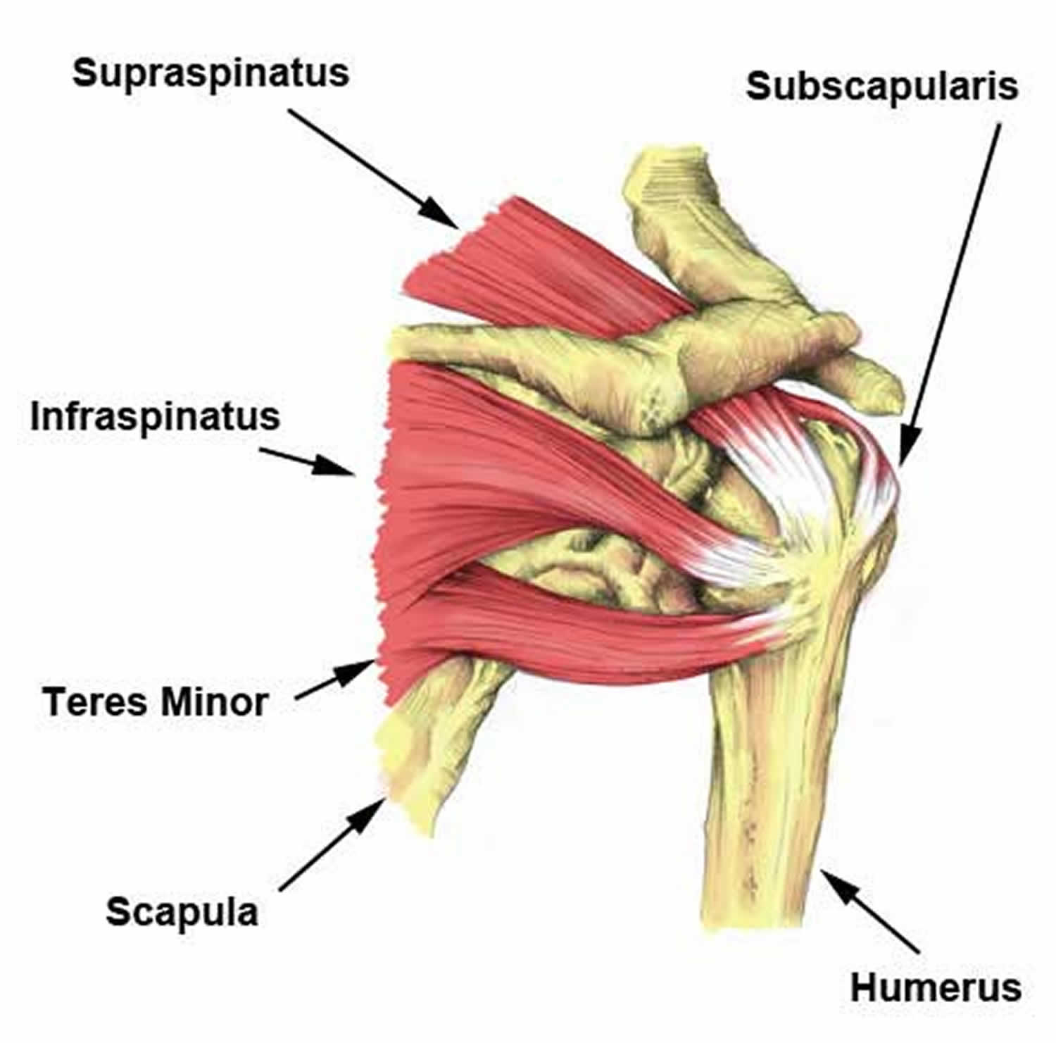
Anterior shoulder pain causes, symptoms, diagnosis & treatment from healthjade.net The bursa is a small sac of fluid that cushions and. Superior glenohumeral ligament and coracohumeral ligament are the primary restraints to posterior acromioclavicular ligament anatomy. Deltoid infraspinatus subscapularis supraspinatus teres major; These four muscles arise from the scapula and insert into the humerus. Posterior — the back of the shoulder medial — the side of the shoulder closest to mid body lateral — the side of the shoulder farthest from mid body proximal — located nearest to the point of attachment or reference, or center of the body You are in the emergency room when a patient is brought in, the loser in a street fight. A muscle contracts to move bones; The muscles and tendons of the rotator cuff form a sleeve around the anterior, superior, and posterior humeral head and glenoid cavity of the shoulder by compressing the glenohumeral joint.
An image depicting shoulder anatomy can be seen below.
Anterior fibers flex & medially rotate arm; Rotator cuff tears, biceps tendon tear at the shoulder. A muscle contracts to move bones; Your rotator cuff consists of the muscles and tendons in your shoulder. Inserts onto navicular tuberosity and first cuneiform. The main function of the triceps is the extension of the elbow joint. It also helps you raise and rotate your arm. You are in the emergency room when a patient is brought in, the loser in a street fight. Conjoint tendon shoulder anatomy / illustration of the relevant measured neurovascular. The tendons of the rotator cuff muscles blend with the joint capsule and form a musculotendinous collar that surrounds the posterior, superior, and anterior aspects of the joint, leaving the inferior aspect unprotected. There are 10 muscles and 11 shoulder tendons related to shoulder mobility. The shoulder anatomy includes the anterior deltoid, lateral deltoid, posterior deltoid, as well as the 4 rotator cuff. The tendons are the attachment of the muscle to the bone.
Posterior to the scapula and inferior to the scapular spine, the infraspinatus tendon inserts on the middle facet of the greater tuberosity, overlapping the posterior aspect of the supraspinatus tendon shoulder tendon anatomy. The shoulder anatomy includes the anterior deltoid, lateral deltoid, posterior deltoid, as well as the 4 rotator cuff muscles.


0 Komentar